Cardiac imaging available with new portable device
Engineers and physicians have developed a wearable ultrasound device that can assess both the structure and function of the human heart. The portable device, which is roughly the size of a postage stamp, can be worn for up to 24 hours and works even during strenuous exercise.
The goal is to make ultrasound more accessible to a larger population, said Sheng Xu, a professor of nanoengineering at the University of California San Diego, who is leading the project. Currently, echocardiograms– ultrasound examinations for the heart– require highly trained technicians and bulky devices.
Thanks to custom AI algorithms, the device is capable of measuring how much blood the heart is pumping. This is important because the heart not pumping enough blood is at the root of most cardiovascular diseases. And issues with heart function often manifest only when the body is in motion. The work is described in the Jan. 25 issue of the journal Nature.
In comparison, existing non-invasive methods have limited sampling capabilities and provide limited data. The wearable technology developed by Xu’s team enables safe, non-invasive and high-quality cardiac imaging, resulting in images with high spatial resolution, temporal resolution and contrast. “It also minimizes patient discomfort and overcomes some limitations of noninvasive technologies such as CT and PET, which could expose patients to radiation,” said Hao Huang, a PhD student in the Xu group at UC San Diego.
The unique design of the sensor makes it ideal for bodies in motion. “The device can be attached to the chest with minimal constraint to the subjects’ movement, even providing a continuous recording of cardiac activities before, during and after exercise,” said Xiaoxiang Gao, a postdoctoral researcher in the Xu group at UC San Diego.
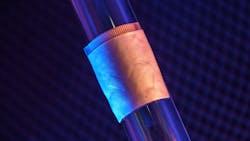
The new system gathers information through a wearable patch as soft as human skin, designed for optimal adherence. The patch measures 1.9 cm (L) x 2.2 cm (W) x 0.09 cm (T), about the size of a postage stamp. It sends and receives the ultrasound waves which are used to generate a constant stream of images of the structure of the heart in real time. This ultrasound patch is soft and stretchable, and it adheres well to human skin, even during exercise.
The system can examine the left ventricle of the heart in separate bi-plane views using ultrasound, generating more clinically useful images than were previously available. As a use case, the team demonstrated imaging of the heart during exercise, which is not possible with the rigid, cumbersome equipment used in clinical settings.
Xu’s team developed an algorithm to facilitate continuous, AI-assisted automatic processing.
“A deep learning model automatically segments the shape of the left ventricle from the continuous image recording, extracting its volume frame-by-frame and yielding waveforms to measure stroke volume, cardiac output and ejection fraction,” said Mohan Li, a master’s student in the Xu group at UC San Diego.
“Specifically, the AI component involves a deep learning model for image segmentation, an algorithm for heart volume calculation, and a data imputation algorithm,” said Ruixiang Qi, a master’s student in the Xu group at UC San Diego. “We use this machine learning model to calculate the heart volume based on the shape and area of the left ventricle segmentation. The imaging-segmentation deep learning model is the first to be functionalized in wearable ultrasound devices. It enables the device to provide accurate and continuous waveforms of key cardiac indices in different physical states, including static and after exercise, which has never been achieved before.”
In the current iteration, the patch is connected through cables to a computer, which can download the data automatically while the patch is still on. The team has developed a wireless circuit for the patch, which will be covered in a forthcoming publication.