The new age of visual inspection

Visual inspection of surgical instruments following decontamination is a universal requirement for surgical instrument reprocessing. Despite the ubiquity of the practice, visual inspection technology has been slow to move into the digital age. In most sterile processing departments lighted desktop magnifying lenses are a common sight. However, the time has come for sterile processing departments to adopt better technology in order to give their technicians the best possible resources to keep patients safe.
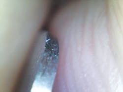
The most significant difference between desktop magnifiers and digital microscopy is the degree of magnification. Most desktop magnifiers are only capable of 2x-4x magnification, depending on the model. Digital microscopy can magnify upwards of 250x. This crack in a tungsten carbide mayo scissor is a perfect example of the need for a higher degree of magnification (see Figure 1).

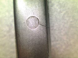
To the naked eye, this crack appeared to be only a small scratch in the finish of the instrument. However, upon closer examination this critical failure in the instrument integrity was found. Such failures are not as rare as some might believe. Even new instruments have been found to have very subtle flaws. This unfinished edge on a malleable ribbon retractor was found by a technician who noticed something felt wrong while opening a set of new instruments (see Figure 2).
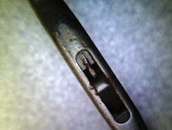
These pictures represent another great advantage of digital microscopy over desktop magnification: the ability to record instrument failures as training opportunities. Anyone who has ever trained in sterile processing can relate to the difficulty in explaining the how’s and why’s of subtle instrument failures. Pictures, on the other hand, can be used very effectively to explain where trouble spots are and what failures look like to new technicians, such as this cracked box lock (see Figure 3).
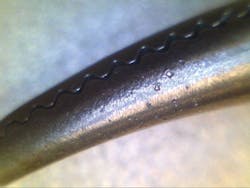
Imagine how difficult this article would have been to understand without the pictures. This feature can be especially helpful when integrated with a digital tracking system. Most tracking systems will allow you to attach pictures and videos to the description of instruments. Such options can help close the gap between experienced SPD technicians and newer technicians. This can be particularly helpful with instruments which have small, difficult to clean areas such as this endoscopic nasal grasper (see Figure 4).
Additionally, pictures of instrument wear and tear can be used to more effectively communicate the toll that poor point-of-use care can have on instruments to the OR. For example, this picture clearly displays the effect that dried saline can have on instruments (see Figure 5).
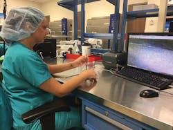
At Baptist Health Medical Center in Conway, AR, we decided to exclusively use the Healthmark MICR007 USB Mircoscope to assist our technicians with visual inspection. The results have been so positive that the use of the digital microscope is now being standardized throughout our nine-hospital system. Additionally, the low cost of entry ($300 per microscope) allows for departments to provide improved visual inspection without investing large amounts of capital.
Subtle instrument failures can have dramatic impact on surgical outcomes. Broken scissors can leave behind fragments inside patients, unfinished edges on a ribbon retractor can result in torn vessels, and hidden bioburden can result in infections. Failures in sterile processing have a direct impact on patient outcomes and it is critical that leadership give technicians every tool necessary to perform at the highest level.